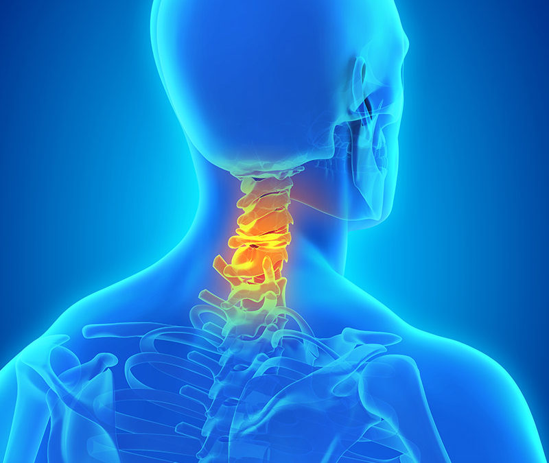
Back Of Neck Anatomy : Primary Neck Cancer Anatomy. Level ii upper internal jugular nodes, posterior to the back of the submandibular salivary gland, anterior to the back of the sternocleidomastoid. Each nerve provides sensation to a specific area of the body called a dermatome. The neck is unique in that it supports the weight of your head (10 to 11 pounds) and allows a variety of head/neck movement, such as. Neck anatomy explained the neck begins at the base of the skull and connects to the thoracic spine (the upper back). Working in pairs on the left and right sides of the body, these muscles.
Below the neck, holding the tooth into the bone, is the root of the tooth. It consists of two major parts: Vertebral, visceral and two vascular compartments. Lymph travels through the lymphatic capillaries, which join together at lymph nodes so that the lymph can be filtered and passed on to larger lymphatic vessels. Neck anatomy explained the neck begins at the base of the skull and connects to the thoracic spine (the upper back).
A Literal Pain In The Neck The Center from www.thecenteroregon.com See anatomy of the head and neck stock video clips. The lymphatic system parallels the cardiovascular system (see the images below). Muscle anatomy coloring sheets 12 photos of the muscle anatomy coloring sheets free muscle anatomy coloring sheets, muscle anatomy coloring pages, muscle anatomy coloring pages free, muscle anatomy coloring sheets, human muscles, free muscle anatomy coloring sheets. Back of neck anatomy lymph / lymph nodes of the head neck and arm kenhub. The structures of the human neck are anatomically grouped into four compartments; In the back, the neck reaches the c7 vertebra. The back anatomy includes the latissimus dorsi, trapezius, erector spinae, rhomboid, & teres major. Related posts of muscle anatomy back of neck muscle anatomy coloring sheets.
Neck anatomy explained the neck begins at the base of the skull and connects to the thoracic spine (the upper back).
The neurocranium (cranial vault) and the viscerocranium (facial skeleton). The back of the neck is mostly comprised of muscles, as well as the spine. Neck anatomy explained the neck begins at the base of the skull and connects to the thoracic spine (the upper back). The majority of these nerves control the functions of the upper extremities and allow you to feel your arms, shoulder, and back of your head. The anterior triangle of the neck is made by the anterior border of the sternocleidomastoid muscle, the inferior border of the mandible and the midline of the neck. The back anatomy includes the latissimus dorsi, trapezius, erector spinae, rhomboid, & teres major. They move the head in every direction, pulling the skull and jaw towards the shoulders, spine, and scapula. Neck anatomy explained the neck begins at the base of the skull and connects to the thoracic spine (the upper back). The neck is the part of the body on many vertebrates that connects the head with the torso and provides the mobility and movements of the head. Rarely, neck pain can be a symptom of a more serious problem. Instant anatomy is a specialised web site for you to learn all about human anatomy of the body with diagrams, podcasts and revision questions. It is made up of bones, discs, muscles, ligaments, nerves and tendons. The occipital bone surrounds a large opening known as the foramen magnum.
The anterior, and the posterior, triangles of the neck. Back of neck anatomy lymph / lymph nodes of the head neck and arm kenhub. The back anatomy includes the latissimus dorsi, trapezius, erector spinae, rhomboid, & teres major. The back comprises the spine and spinal nerves, as well as several different muscle groups. The neck is unique in that it supports the weight of your head (10 to 11 pounds) and allows a variety of head/neck movement, such as.
Female Back And Neck Anatomy Illustration Stock Image F026 5709 Science Photo Library from media.sciencephoto.com The neck begins at the lower edge of the jaw and the occipital bone, which is the base of the skull. The lymphatic system parallels the cardiovascular system (see the images below). These two ligaments connect and support the spine from the neck to the lower. Overview of head and neck tumors. The occipital bone is the only bone in your head that connects with your cervical spine (neck). Instant anatomy is a specialised web site for you to learn all about human anatomy of the body with diagrams, podcasts and revision questions. Related posts of muscle anatomy back of neck muscle anatomy coloring sheets. Think of it like a jigsaw puzzle, all the pieces fit in together and are required to get the full picture as to how it works.
Then it extends to the clavicles and the sternum in front.
They move the head in every direction, pulling the skull and jaw towards the shoulders, spine, and scapula. Learn everything about the neck anatomy with this topic page. It consists of two major parts: The back comprises the spine and spinal nerves, as well as several different muscle groups. In the back, the neck reaches the c7 vertebra. In addition, in this region we also find the major cranial and spinal nerves that connect the central nervous system to the organs, skin, and muscles of the head and neck. The back of the neck is mostly comprised of muscles, as well as the spine. Below the neck, holding the tooth into the bone, is the root of the tooth. Back of neck anatomy lymph / lymph nodes of the head neck and arm kenhub. Instant anatomy is a specialised web site for you to learn all about human anatomy of the body with diagrams, podcasts and revision questions. The lymphatic system parallels the cardiovascular system (see the images below). Muscle head anatomy vocal organ diagram female neck anatomy neck wireframe head neck human anatomy head artery anatomy face pharynx vector neck degree head anatomy 3d. The neck is unique in that it supports the weight of your head (10 to 11 pounds) and allows a variety of head/neck movement, such as.
When most people mention their back, what they are actually referring to is their spine. From the sides and the back of the neck, the splenius capitis inserts onto the head region, and the splenius cervicis extends onto the. The occipital bone is the only bone in your head that connects with your cervical spine (neck). The top of the cervical spine connects to the skull, and the bottom connects to the upper back at about shoulder level. Back of neck anatomy lymph / lymph nodes of the head neck and arm kenhub.
Anatomy Of Back Of Neck Anatomy Drawing Diagram from media.istockphoto.com Overview of head and neck tumors. The skull is a strong, bony capsule that rests on the neck and encloses the brain. The splenius muscles originate at the midline and run laterally and superiorly to their insertions. The neck is unique in that it supports the weight of your head (10 to 11 pounds) and allows a variety of head/neck movement, such as. The neurocranium (cranial vault) and the viscerocranium (facial skeleton). The structures of the human neck are anatomically grouped into four compartments; It is made up of bones, discs, muscles, ligaments, nerves and tendons. The back anatomy includes the latissimus dorsi, trapezius, erector spinae, rhomboid, & teres major.
Two of the main ligaments in the back are the anterior longitudinal ligament and the posterior longitudinal ligament.
The majority of these nerves control the functions of the upper extremities and allow you to feel your arms, shoulder, and back of your head. Learn everything about the neck anatomy with this topic page. Below the neck, holding the tooth into the bone, is the root of the tooth. See human neck anatomy stock video clips. In the back, the neck reaches the c7 vertebra. From the sides and the back of the neck, the splenius capitis inserts onto the head region, and the splenius cervicis extends onto the. Level ii upper internal jugular nodes, posterior to the back of the submandibular salivary gland, anterior to the back of the sternocleidomastoid. It consists of two major parts: Working in pairs on the left and right sides of the body, these muscles. Cervical spine anatomy (neck) the cervical spine, your neck, is a complex structure making up the first region of the spinal column starting immediately below the skull and ending at the first thoracic vertebra. The top of the cervical spine connects to the skull, and the bottom connects to the upper back at about shoulder level. Related posts of muscle anatomy back of neck muscle anatomy coloring sheets. Each nerve provides sensation to a specific area of the body called a dermatome.
Share :
Post a Comment
for "Back Of Neck Anatomy : Primary Neck Cancer Anatomy"


Post a Comment for "Back Of Neck Anatomy : Primary Neck Cancer Anatomy"